Key Takeaways
-
Position electrode pads proximal and distal to the pain site rather than directly on it to ensure current traverses affected tissue and engages relevant nerve fibers effectively.
-
Maintain at least one inch of separation between pads to ensure adequate current penetration into deeper tissues rather than taking the path of least resistance on skin surface.
-
Avoid high-risk anatomical zones including anterior cervical region, head/face, across the chest, broken skin, and areas near implanted electronic devices like pacemakers.
-
Align pad placement with dermatome patterns for nerve pain conditions such as radiculopathy, allowing current to target specific nerve root distribution for more substantial relief.
-
Document exact electrode placement coordinates including anatomical landmarks and inter-electrode distance to replicate effective treatments and ensure clinical consistency across sessions.
-
Prepare skin properly by cleaning with mild soap and water, ensuring complete dryness, and avoiding lotions or oils before pad application to maximize current delivery and pad adhesion.
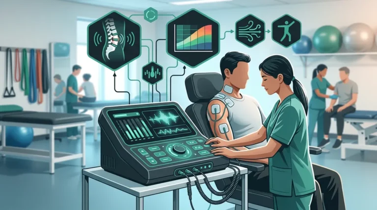
Proper TENS unit pads placement is one of the most consequential decisions a clinician makes when administering electrotherapy. Whether you are managing post-injury rehabilitation in a physical therapy clinic, supporting spinal care in a chiropractic practice, or treating soft tissue damage from an auto accident, electrode positioning directly determines the quality of pain relief your patients experience. Misplaced pads reduce therapeutic effectiveness, may cause patient discomfort, and can undermine confidence in the treatment protocol.
For physical therapy providers, chiropractors, and auto accident injury clinics in 2026, understanding the clinical nuances of electrode placement is not optional — it is fundamental to delivering measurable outcomes. This guide presents ten evidence-based placement tips designed to help clinicians optimize their electrotherapy protocols and improve patient satisfaction. As noted by the Cleveland Clinic, TENS therapy is a well-established non-pharmacological pain management option that works most effectively when electrodes are positioned correctly relative to the targeted nerve pathways.
Why TENS Unit Pads Placement Matters in Clinical Settings
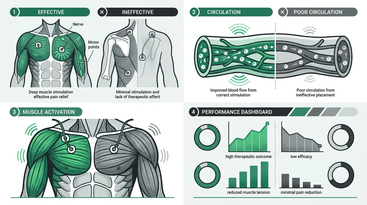
The therapeutic effect of a TENS unit depends on delivering electrical current through specific tissues to modulate pain signals. When pads are placed accurately, the current reaches the intended sensory nerves, activating the pain gate mechanism or stimulating endorphin release. Incorrect placement, however, means the current may bypass the target entirely or stimulate unintended tissues. For clinics managing high volumes of patients with diverse pain conditions, a standardized yet adaptable placement protocol is essential.
Understanding what TENS unit pads actually do for pain relief provides clinicians with the mechanistic foundation to make informed placement decisions. For clinics seeking a deeper technical overview, this electrotherapy electrodes clinical guide for providers is an excellent resource.

10 Clinical Tips for Optimal TENS Unit Pads Placement
1. Position Pads Around, Not Directly On, the Pain Site
A foundational principle in electrotherapy is that electrodes should flank the area of pain rather than sit directly on it. Placing one pad proximal and one distal to the pain site allows the electrical current to traverse the affected tissue, engaging the relevant nerve fibers effectively. This technique is especially useful for localized joint pain, nerve root irritation, and soft tissue injuries common in auto accident cases.
2. Maintain a Minimum One-Inch Pad Separation
Pads placed too close together cause the current to take the path of least resistance between the electrodes on the skin surface, rather than penetrating deeper tissues. Clinicians should maintain at least one inch of separation between pads to ensure adequate current dispersion. For larger muscle groups such as the lumbar paraspinals or quadriceps, wider spacing is appropriate to encompass the target area effectively.
3. Align Placement with Dermatome Patterns for Nerve Pain
For patients presenting with radiculopathy or referred pain — conditions frequently encountered after spinal injuries or auto accidents — aligning pad placement with the relevant dermatome is clinically significant. Placing electrodes along the dermatome path allows the current to target the specific nerve root distribution, which may provide more substantial relief than placement at the pain site alone. Nerve stimulators in electrotherapy follow similar anatomical principles that chiropractors and physical therapists can apply directly.
4. Apply Pads Bilaterally for Symmetrical Pain Conditions
For conditions such as bilateral lumbar pain, sacroiliac dysfunction, or symmetrical joint degeneration, bilateral pad placement can deliver more comprehensive relief. Placing one electrode on each side of the spine — or each side of a symptomatic joint — allows the electrical current to cover a broader treatment area. This approach is particularly effective in chiropractic settings where spinal alignment and bilateral muscle symmetry are clinical priorities.
5. Avoid High-Risk Anatomical Zones
Certain anatomical zones are contraindicated for electrode placement regardless of the pain location. These include the anterior cervical region (front of the neck), the head and face, across the chest (particularly transcardiac placement), broken or irritated skin, and areas near implanted electronic devices. Clinicians must screen patients for pacemakers, spinal cord stimulators, and other implants before initiating TENS therapy. The FDA provides guidance on safe use of non-opioid pain therapies, including electrical stimulation devices.
| Contraindicated Zone | Reason for Avoidance | Clinical Alternative |
|---|---|---|
| Anterior cervical (front of neck) | Risk of laryngospasm or blood pressure changes | Posterior cervical or trapezius placement |
| Across the chest | Potential for cardiac interference | Unilateral thoracic or upper back placement |
| Head and face | Risk of seizure or unintended nerve stimulation | Cervical or occipital region (with caution) |
| Over broken or irritated skin | Increased burn risk and patient discomfort | Proximal or distal to the affected area |
| Near implanted electronic devices | Interference with device function | Contraindicated — consult device manufacturer |
6. Use Conductive Garments for Consistent Coverage
Traditional adhesive electrodes can be difficult to position consistently, particularly for patients using TENS at home between clinic visits. Conductive garments offer a practical solution by integrating electrode surfaces directly into wearable garments, ensuring reproducible placement every session. For patients managing chronic low back pain or recovering from workplace injuries, this consistency translates into more predictable therapeutic outcomes. TheraKnit Garments represent an advanced option in this category, providing uniform current distribution across larger treatment areas.
7. Calibrate Placement Based on Stimulation Frequency and Mode
TENS units offer multiple frequency settings — high-frequency (conventional TENS) and low-frequency (acupuncture-like TENS) — each with distinct physiological effects. High-frequency stimulation (80–150 Hz) targets sensory nerve fibers and is effective for acute pain, while low-frequency stimulation (1–10 Hz) targets motor nerve fibers and promotes endorphin release for chronic pain. Pad placement should be adapted accordingly: high-frequency modes benefit from direct peripheral nerve coverage, while low-frequency modes may be more effective when placed over motor points or acupuncture-adjacent sites.
| TENS Mode | Frequency Range | Recommended Placement Strategy | Best Suited For |
|---|---|---|---|
| Conventional (High-Frequency) | 80–150 Hz | Around the pain site, over sensory nerves | Acute pain, post-injury |
| Acupuncture-Like (Low-Frequency) | 1–10 Hz | Over motor points or trigger points | Chronic pain, muscle tension |
| Burst Mode | Intermittent bursts | Combination of sensory and motor point targeting | Mixed acute and chronic presentations |
8. Apply Specific Placement Protocols for Common Clinical Conditions
Standardizing placement protocols for frequently treated conditions improves clinical efficiency and patient outcomes. The following list outlines placement strategies for conditions commonly encountered in chiropractic, physical therapy, and auto accident injury practices:
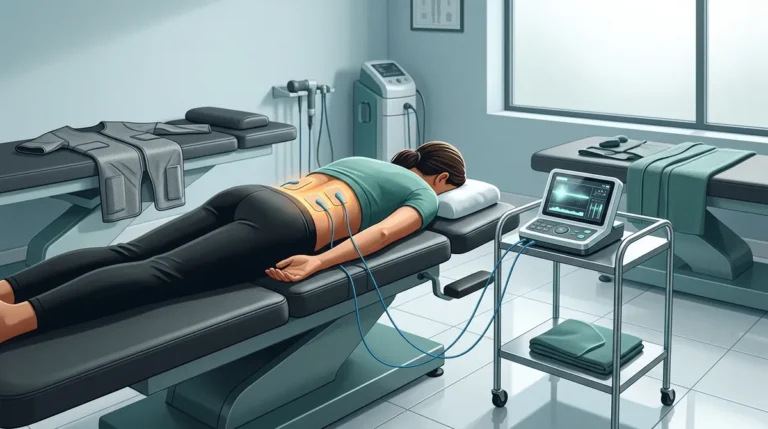
- Lumbar back pain: Place one electrode on each side of the lumbar spine at the level of pain, approximately two inches from the midline. A second pair may be positioned over the gluteal region for radiating pain.
- Cervical pain and whiplash: Position electrodes on the posterior cervical muscles bilaterally, avoiding the anterior neck. For trapezius involvement, extend placement to the upper trapezius region.
- Knee pain: Place electrodes medially and laterally around the joint, or above and below the patella for comprehensive coverage.
- Shoulder pain: Position one electrode over the deltoid and one over the posterior rotator cuff, adjusting based on the specific structure involved.
- Sciatic nerve pain: Follow the sciatic nerve path with electrodes placed at the lumbar origin and along the posterior thigh toward the knee.
For providers treating auto accident patients, proper cervical and lumbar electrode placement is particularly critical. Reviewing available TENS unit devices alongside placement protocols ensures the right equipment is matched to the clinical need.
9. Ensure Optimal Skin Preparation Before Pad Application
Skin condition at the electrode site significantly affects current delivery and pad adhesion. Oily, dirty, or excessively dry skin increases impedance, reducing the current reaching the target tissue. Clinicians should instruct patients — and clinical staff — to clean the skin with mild soap and water before applying pads, allow the skin to dry completely, and avoid applying lotions or oils to the treatment area. Proper skin preparation also extends pad life, reducing supply costs over time. Essential medical supplies for electrotherapy clinics include quality electrode gels and skin preparation wipes as standard components of a well-stocked treatment suite.
10. Document Placement Coordinates for Treatment Consistency
Clinical consistency is a hallmark of effective electrotherapy programs. Documenting exact electrode placement coordinates — including anatomical landmarks, inter-electrode distance, and patient-reported response — allows clinicians to replicate effective treatments and make precise adjustments when outcomes are suboptimal. For practices managing large patient volumes, standardized placement documentation also supports billing accuracy and compliance with insurance requirements, including workers’ compensation and auto accident claims.
According to the National Institutes of Health, chronic pain affects a substantial portion of U.S. adults, underscoring the clinical importance of precise, reproducible electrotherapy protocols. The CDC also recognizes non-opioid therapies, including TENS, as a preferred pain management strategy — making proper electrode placement a public health-relevant clinical skill.
Common TENS Pad Placement Errors and How to Avoid Them
Even experienced clinicians can fall into placement habits that reduce treatment efficacy. The following table identifies common errors and their clinical corrections:
| Common Error | Clinical Impact | Recommended Correction |
|---|---|---|
| Pads placed directly on the pain epicenter | Reduced current penetration to target tissue | Flank the pain site with proximal and distal placement |
| Insufficient pad spacing | Current bypasses deep tissue, stays superficial | Maintain minimum one-inch separation; wider for large muscles |
| Placement over scar tissue or bony prominences | Poor conductivity, patient discomfort | Reposition to adjacent soft tissue areas |
| Failure to rotate pad placement sites | Skin irritation, reduced adhesion quality | Rotate placement site with each session |
Integrating Placement Best Practices into Your Clinical Workflow
Implementing a structured approach to TENS unit pads placement requires both staff education and the right equipment infrastructure. Physical therapy clinics benefit from laminated placement reference guides at each treatment station, while chiropractic practices may integrate placement protocols into their electronic health record (EHR) templates. Auto accident injury clinics, managing patients through insurance claim processes, benefit from detailed placement documentation to support clinical justification in billing.
Liberty Medical Solutions supports healthcare providers with customized electrotherapy solutions — including TENS units, conductive garments, and back braces — designed to integrate seamlessly into clinical workflows. The company also works with commercial PPO/POS plans, workers’ compensation, and auto accident insurance claims, making it a practical partner for multi-specialty practices. For clinicians looking to expand their electrotherapy capabilities, exploring the full range of available electrotherapy products is a logical next step. Providers interested in clinical-grade electrode options can also review what makes the best TENS machine for clinical use to align equipment selection with placement best practices.
Selecting the Right Electrode for the Placement Site
Pad geometry plays a meaningful role in treatment outcomes. Smaller electrodes concentrate current density, making them appropriate for precise, localized targets such as trigger points or small joints. Larger electrodes distribute current over a broader surface area, which is preferable for large muscle groups or bilateral spinal applications. Matching electrode size to the anatomical target and treatment goal is an often-overlooked dimension of effective TENS unit pads placement.
- Small electrodes (1″–2″): Best for localized nerve points, small joints, and trigger point therapy
- Medium electrodes (2″–3″): Suitable for extremity muscles, shoulder, and forearm applications
- Large electrodes (3″–4″ and above): Recommended for lumbar, thoracic, and large quadriceps applications
- Conductive garments: Ideal for full-region coverage, at-home consistency, and patients with limited dexterity
Conclusion
Mastering TENS unit pads placement is a foundational competency for any clinical practice using electrotherapy as part of its pain management protocol. From understanding dermatome-based positioning to avoiding contraindicated zones and documenting placement coordinates for reproducibility, each of these ten tips contributes directly to improved patient outcomes and clinical efficiency. For physical therapy clinics, chiropractic practices, and auto accident injury providers in 2026, a precision-focused approach to electrode placement is not just best practice — it is a competitive clinical advantage.
If your practice is ready to elevate its electrotherapy program with the right devices, electrodes, and clinical support, reach out to our team at Liberty Medical Solutions to discuss customized solutions tailored to your patient population and insurance billing needs.
FAQs
Q: Where should TENS unit pads be placed for lower back pain?
A: For lower back pain, electrodes should be positioned on either side of the lumbar spine at the level of discomfort, approximately two inches from the midline. A second pair of pads may be placed over the gluteal region if pain radiates downward. Always avoid placement directly over the spine or on broken skin.
Q: Can TENS unit pads be placed on the neck for whiplash injuries?
A: Yes, TENS electrodes can be placed on the posterior cervical muscles to address whiplash-related pain. Placement on the front of the neck is strictly contraindicated due to risks of laryngospasm and blood pressure interference. Clinicians should position pads on the posterior cervical and upper trapezius region for safe and effective treatment.
Q: How far apart should TENS pads be placed from each other?
A: A minimum of one inch of separation between electrodes is recommended to ensure the electrical current penetrates into deeper tissues rather than traveling superficially between pads. For larger muscle groups such as the lumbar paraspinals or quadriceps, wider spacing of two to four inches is appropriate for broader current distribution.
Q: Are conductive garments an effective alternative to standard TENS electrode pads?
A: Conductive garments provide a clinically effective alternative, particularly for patients requiring consistent at-home electrode placement between clinic visits. They integrate electrode surfaces directly into wearable material, ensuring reproducible positioning with each use. This consistency supports more predictable therapeutic outcomes compared to self-applied adhesive pads.
Q: What areas of the body are contraindicated for TENS unit pad placement?
A: Contraindicated areas include the anterior cervical region (front of the neck), across the chest, the head and face, over broken or irritated skin, and near any implanted electronic device such as a pacemaker or spinal cord stimulator. Clinicians must conduct a thorough patient screening prior to initiating TENS therapy to identify any relevant contraindications.